Cells, Water, Balloons and Sponges
Intro
Richard Feinman.
My old friend Fred Sachs is a well-known biophysicist at SUNY Buffalo. Among other accomplishments, he discovered mechanosensitive ion channels. These receptors respond to physical pressure and so they provide a mechanism for cells to transmit the information about mechanical stress by converting the input to an electrical signal. Fred asked me once in conversation if I knew what would happen if you put a living cell (with all its dissolved ions, proteins and metabolites) in distilled water. I know that the cell is not “a bag of water” but my knowledge is not deep on the subject. We all know about osmosis in a rough sort of way — water will pass through a membrane into a solution of higher concentration until the concentrations even out. So, my first reaction was that water would keep moving into the cell until it exploded. It turns out this is not true and he explained what his research had identified. It turns out that many more established researchers also didn’t get it right. If you can believe it, established scientists resisted changing their old ideas in light of new experiments. Here’s the story.
Cells, Water, Balloons and Sponges.
Frederick Sachs
It’s true. Most living cells in higher organisms will not explode in distilled water. The simple idea of osmosis as we learn it in school is not really applicable. Most cells are not like a balloon filled with liquid and do not really depend substantially on the cell membrane to keep them intact. Most living cells are more like sponges. They take up water when placed in a bath of low concentration but they have a network of internal structures that provides its own resistance, that is, the whole network can contract or expand (Figure 1). Chemically, such a structure is referred to as a gel — mostly water but because of the structure, significant physical strength. (Think of how much water you add to the teaspoon of powder to make Jell-O).
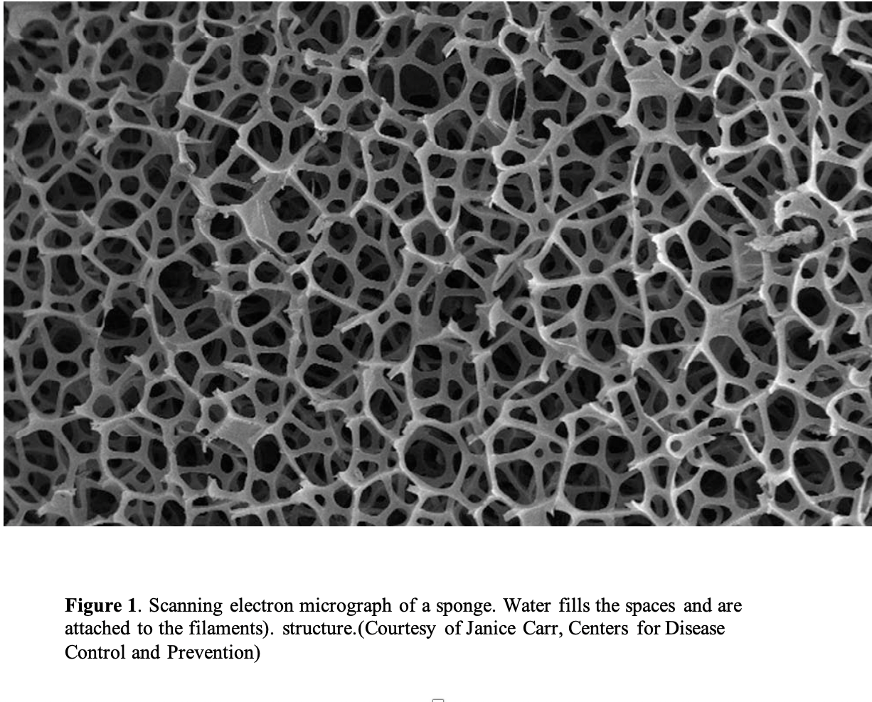
The sponge-like network of the cell is referred to as its cytoskeleton and it represents a big player in the life of the cell. Figure 2 shows the network of actin (a contractile protein), one of the proteins that provides the structure of the cytoskeleton. Cross-linking creates an elaborate structure which is attached to the cell membrane but, unlike what we thought from old ideas, it’s not the membrane that keeps the cell together but rather the strength of the multiple interactions of the cross-linked cytoskeleton. Some simpler cells, like red blood cells, behave more like a balloon — red cells will explode in distilled water. Bacteria and plants have to have a physical cell wall but this is obviously limited and less dynamic.

If you think about it, when a cell divides you end up with two cells. In many organs, the daughter cells are almost all the same size. How do these cells know how big to get or when to stop growing? The living cell does not contain anything like a ruler to measure its size. The cytoskeleton is a built-in control on the size of the cell.
So, how does a sponge swell? The fibers of a sponge are wettable, or hydrophillic, meaning that they stick to water molecules. (Looking ahead the cytoskeleton is made of proteins which associate with water as well). Now, initially, watersticks to the polymers that make up the sponge. After a few water molecules stick, additional water flowing into the sponge looks like bulk water. The water continues to flow in because there is room for it. The second law of thermodynamics tells us things will always try to spread out. At some point, the sponge stops swelling and reaches equilibrium because transferring water into the sponge means you have to make room for it. To do that you have to stretch the polymer threads that make up the network.
That requires mechanical work. Equilibrium is reached when the entropic energy (spreading out) for water diffusion into the sponge equals the hydrostatic pressure of the water in the free volume.
Predictions and Experiment
How can we test this theory? Well, if the cell were like a balloon, or like a football, it would get stiffer as we “inflate” it. To make measurements on the stiffness of single cells, we needed a rather sophisticated set-up. In the end, we used an atomic force microscope that could give readings of atomic level forces. The discussion in this post is a summary of our published review and the AFM study. I’ll finish the post by citing from those reports what happened next:
“I asked my postdoc, Dr. Chiara Spagnoli, who was fluent in atomic force microscopy (AFM) to see how stiff cells become during hypoosmotic swelling, postulating the dogma that they should get stiffer as they inflate. However, after months of testing and many controls, she found that the cells stayed the same or got softer with swelling. After much agonizing, we realized that the behavior was much like how sponges behave,”
A result from Dr. Spagnoli’s experiment is shown in Figure 3.
“The cell height and Young’s modulus [stiffness] of a rat astrocyte [glial cell] in response to a hypotonic challenge. As expected, the cell swelled …. And the Young’s modulus decreased as the cell swelled, and rather than becoming stiff as one might expect of an inflated vesicle, i.e., the cell became softer.”
